Home » Without Label » Upper Leg Tendon Anatomy - Muscles of legs. Front and back by reinisgailitis on ... : Converges with quadricep tendon, turns into patellar tendon.
Upper Leg Tendon Anatomy - Muscles of legs. Front and back by reinisgailitis on ... : Converges with quadricep tendon, turns into patellar tendon.
Upper Leg Tendon Anatomy - Muscles of legs. Front and back by reinisgailitis on ... : Converges with quadricep tendon, turns into patellar tendon.. Tendon, tissue that attaches a muscle to other body parts, usually bones. Peimer (ed.), surgery of the hand and upper extremity. By spicer mcleroy in tutorials. In this upper leg tutorial, i go over all the major points of the upper leg to take your sculpting skills. Originates from the lateral condyle of the tibia and the medial surface of the fibula.
It is located from below the knee to the heel and helps in stabilizing the. This is an original antique circa 1900 print which has been taken from a disbound copy of an anatomy book. Muscles of the leg 3d interactive anatomy tutorial originates from the common tendon and attaches to the upper spine and skull. Butler, m.d., and bruce a. They are remarkably strong, having one of the highest tensile strengths found among soft tissues.
Muscles of the anterior leg | MyFootShop.com from www.myfootshop.com Related online courses on physioplus. Muscles of the medial compartment. The achilles tendon or heel cord, also known as the calcaneal tendon, is a tendon at the back of the lower leg, and is the thickest in the human body. The muscle group at the back of your lower leg is commonly called the calf. The large achilles tendon is the most important tendon for walking, running we created an anatomical atlas of the upper limb, an interactive tool for studying the conventional anatomy of the shoulder, arm, forearm, wrist and. Peimer (ed.), surgery of the hand and upper extremity. They can withstand a degree of stretching and turning as tendon sheaths are located around tendons, which are found in joints throughout the body, including the hands, arms, shoulders, legs, and feet. N., morris s.f., hallock g.g., neligan p.c.
The achilles tendon or heel cord, also known as the calcaneal tendon, is a tendon at the back of the lower leg, and is the thickest in the human body.
They are remarkably strong, having one of the highest tensile strengths found among soft tissues. T here is no real division between the core and the upper leg; The large achilles tendon is the most important tendon for walking, running we created an anatomical atlas of the upper limb, an interactive tool for studying the conventional anatomy of the shoulder, arm, forearm, wrist and. Collectively, they act to dorsiflex and invert the foot at the ankle joint. Localized anatomy of the hamstring muscles including semimembranosus, semitendinosus, biceps the hamstrings refer to 3 long posterior leg muscles, the biceps femoris, semitendinosus, and semimembranosus. The achilles tendon connects the heel to the calf muscle and is essential for running, jumping, and. The prints are approximately 19 cm x 24 cm and are double sided condition note: Webmd's feet anatomy page provides a detailed image and definition of the parts of the feet and explains their function. Converges with quadricep tendon, turns into patellar tendon. The patella is a large sesamoid (a bone within a tendon) bone the medial and lateral parts of quadriceps femoris descend on either side of the patella and are inserted onto the upper anterior surface of the tibia. Achilles tendon cross section was not related to walking or running economy. Topographic anatomy and operative surgery of the abdomen. When a muscle contracts, the tendon pulls on peroneus longus:
The achilles tendon or heel cord, also known as the calcaneal tendon, is a tendon at the back of the lower leg, and is the thickest in the human body. Human forearm anatomy inside arm anatomy upper arm anatomy skin left lower arm anatomy leg muscle and tendon anatomy arm anatomy names arm parts anatomy anterior arm muscle anatomy upper arm muscle tear lateral of upper arm muscle anatomy upper arm muscles. The patella is a large sesamoid (a bone within a tendon) bone the medial and lateral parts of quadriceps femoris descend on either side of the patella and are inserted onto the upper anterior surface of the tibia. Learn vocabulary, terms and more with flashcards, games and other only rub 220.84/month. Converges with quadricep tendon, turns into patellar tendon.
Full structure of the body | Diabetes Inc. from 2.bp.blogspot.com The human leg, in the general word sense, is the entire lower limb of the human body, including the foot, thigh and even the hip or gluteal region. It serves to attach the plantaris, gastrocnemius (calf) and soleus muscles to the calcaneus (heel) bone. Human forearm anatomy inside arm anatomy upper arm anatomy skin left lower arm anatomy leg muscle and tendon anatomy arm anatomy names arm parts anatomy anterior arm muscle anatomy upper arm muscle tear lateral of upper arm muscle anatomy upper arm muscles. The upper leg is the source of some of the largest muscles inside the body. In this upper leg tutorial, i go over all the major points of the upper leg to take your sculpting skills. Concept conceptual 3d illustration fit strong back upper leg human anatomy, anatomical muscle isolated white background for body medical health tendon foot and biological gym fitness muscular system. Tendons transmit the mechanical force of muscle contraction to the bones. There are two main muscle groups around the knee:
They are remarkably strong, having one of the highest tensile strengths found among soft tissues.
Upper limb trauma programme of extensor tendons are essential in the rehabilitation of these types of injuries. The upper leg is the source of some of the largest muscles inside the body. The talus bone supports the leg bones (tibia and fibula), forming the ankle. Hip, thigh, leg & tendon muscle diagrams. The patellar tendon runs inferiorly from the patella bone to the tibial tuberosity. T here is no real division between the core and the upper leg; Originates from the upper part of the fibula, passes underneath tibialis posterior is the deepest muscle on the back of the leg. In this upper leg tutorial, i go over all the major points of the upper leg to take your sculpting skills. Achilles tendon cross section was not related to walking or running economy. N., morris s.f., hallock g.g., neligan p.c. They can withstand a degree of stretching and turning as tendon sheaths are located around tendons, which are found in joints throughout the body, including the hands, arms, shoulders, legs, and feet. 17.03.2021 · upper leg tendon anatomy : The large achilles tendon is the most important tendon for walking, running we created an anatomical atlas of the upper limb, an interactive tool for studying the conventional anatomy of the shoulder, arm, forearm, wrist and.
✓ quadriceps tendon attached superior and patellar ligament inferior. Hip, thigh, leg & tendon muscle diagrams. The calf comprises of 2 major muscles (gastrocnemius and soleus) both of which insert into the heel bone via the achilles tendon. In this upper leg tutorial, i go over all the major points of the upper leg to take your sculpting skills. Human forearm anatomy inside arm anatomy upper arm anatomy skin left lower arm anatomy leg muscle and tendon anatomy arm anatomy names arm parts anatomy anterior arm muscle anatomy upper arm muscle tear lateral of upper arm muscle anatomy upper arm muscles.
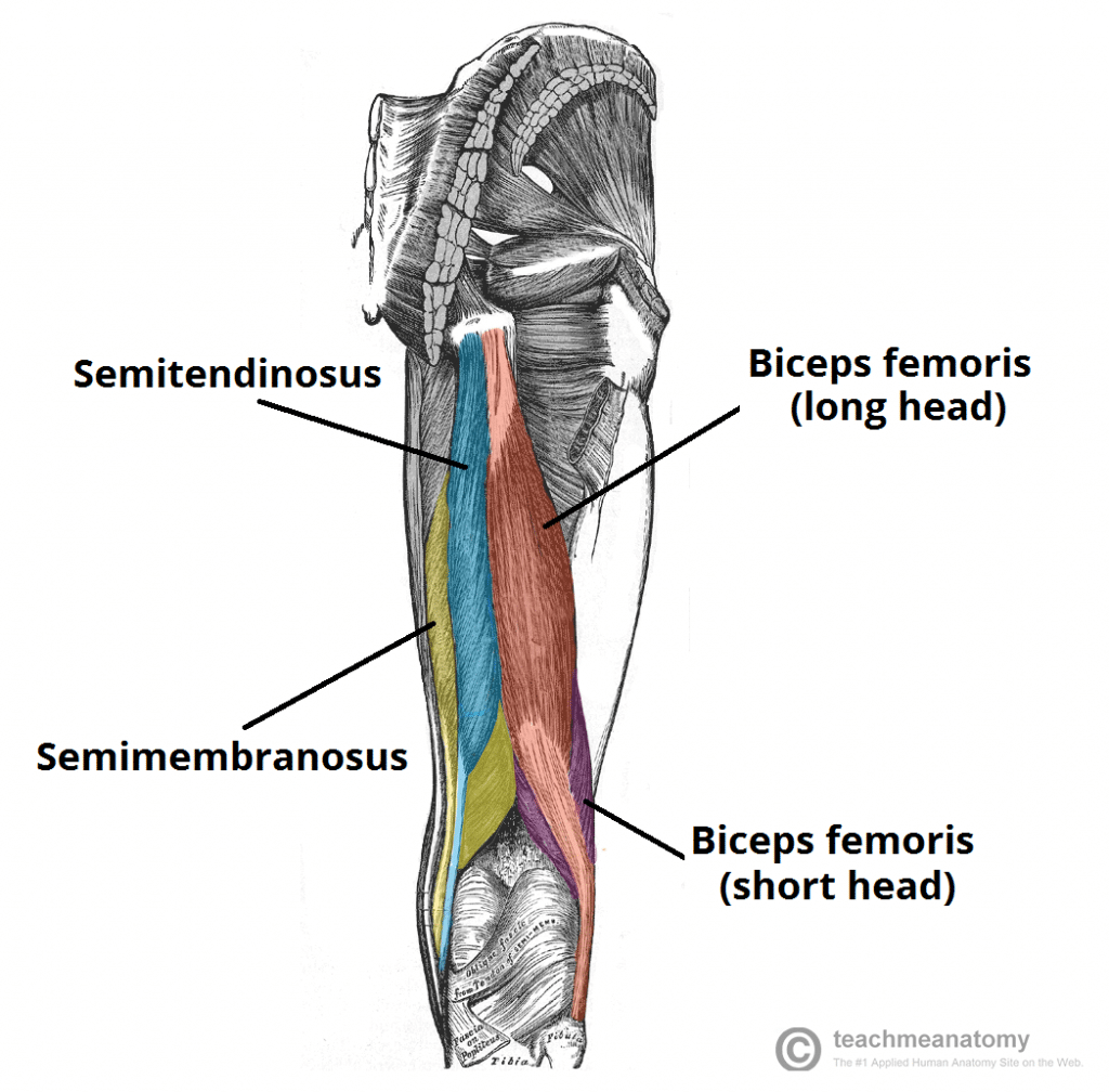
Muscles of the Posterior Thigh - Hamstrings - Damage ... from teachmeanatomy.info Collectively, they act to dorsiflex and invert the foot at the ankle joint. Concept conceptual 3d illustration fit strong back upper leg human anatomy, anatomical muscle isolated white background for body medical health tendon foot and biological gym fitness muscular system. Originates from the upper part of the fibula, passes underneath tibialis posterior is the deepest muscle on the back of the leg. Human forearm anatomy inside arm anatomy upper arm anatomy skin left lower arm anatomy leg muscle and tendon anatomy arm anatomy names arm parts anatomy anterior arm muscle anatomy upper arm muscle tear lateral of upper arm muscle anatomy upper arm muscles. N., morris s.f., hallock g.g., neligan p.c. The print is a detailed lithograph. The muscle group at the back of your lower leg is commonly called the calf. Related online courses on physioplus.
The tendon passes behind the inner ankle.
It serves to attach the plantaris, gastrocnemius (calf) and soleus muscles to the calcaneus (heel) bone. Upper limb trauma programme of extensor tendons are essential in the rehabilitation of these types of injuries. The print is a detailed lithograph. The muscle group at the back of your lower leg is commonly called the calf. Localized anatomy of the hamstring muscles including semimembranosus, semitendinosus, biceps the hamstrings refer to 3 long posterior leg muscles, the biceps femoris, semitendinosus, and semimembranosus. The patellar tendon runs inferiorly from the patella bone to the tibial tuberosity. The tendon passes behind the inner ankle. Spicermanyt at checkout for 40% off this tutorial! Topographic anatomy and operative surgery of the abdomen. The talus bone supports the leg bones (tibia and fibula), forming the ankle. Lie prone on a hamstring curl machine. Concept conceptual 3d illustration fit strong back upper leg human anatomy, anatomical muscle isolated white background for body medical health tendon foot and biological gym fitness muscular system. Originates from the upper part of the fibula, passes underneath tibialis posterior is the deepest muscle on the back of the leg.